On June 14, 2022, with the cooperation of relevant departments of the hospital, Bei Chaoyong, Deputy Director of the Department of Hand Surgery for Extremity Trauma of the Affiliated Hospital of Guilin Medical College, and his team formulated a surgical plan for a patient diagnosed with "left femoral head necrosis (stage II)" using 3D printing guide plate precision positioning technology, and successfully completed a The minimally invasive surgery of "3D printed surgical guide guided by precise core drilling and decompression of the femoral head + bone grafting" was successfully completed.

Mr. Zhang (a pseudonym), 61 years old, was admitted to the Hospital of Guilin Medical College for treatment of left hip pain for one year and severe limping for six months. Prior to his admission, he had visited several hospitals for consultation and was advised by some doctors to have a total hip replacement. However, his will to keep his hip joint was so strong that he made a special trip to the Affiliated Hospital of Guilin Medical College to find Dr. Bei Chaoyong, the chief physician of the Department of Extremity Trauma and Hand Surgery of the hospital.
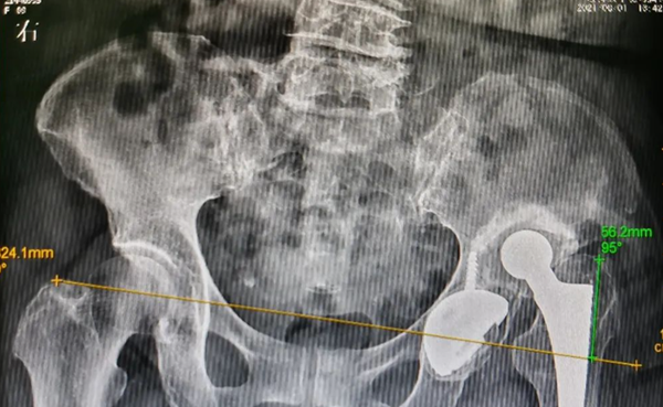
It was understood that Mr. Zhang had a history of surgery for a fractured femoral head from a traumatic injury to his left hip in a car accident 21 years ago. The examination after admission showed that his femoral head was still intact, with several vesicular necrotic changes visible in the central area of the femoral head, no collapse of the femoral head and normal joint space. After a thorough examination, he diagnosed traumatic necrosis of the left femoral head (stage II), which could be treated with hip preservation surgery. However, the patient had four vesicle-like necrotic changes within the femoral head and they were distributed in different parts of the femoral head, with the largest weight-bearing vesicle at the top of the acetabulum about to penetrate the cartilaginous surface of the femoral head.
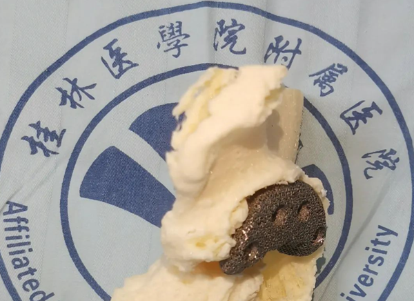
The use of conventional fluoroscopic core decompression surgery was not sufficiently accurate and might not accurately locate the vesicles and could easily penetrate the femoral head and worsen the femoral head necrosis. After repeated discussions in the department, Dr. Pei Chaoyong, the chief surgeon, decided to use a minimally invasive technique of 3D printed surgical guide to perform a left femoral head medullary core drilling and decompression + bone grafting. This technique can precisely locate the location of the femoral head necrosis and design the best angle and direction of needle entry and depth of the medullary core decompression channel to precisely reach the desired necrotic vesicle site, avoiding penetrating the cartilaginous surface of the femoral head. In order to allow for better growth of the femoral head, after discussion among the experts in the department, Dr. Pei Chaoyong, the chief surgeon, and his team decided to use a new medullary decompression rod (bioactive material n-HA/PA66) mixed with autologous iliac bone for bone grafting to fully decompress the pressure within the femoral head, improve the blood supply to the femoral head and prevent the femoral head from collapsing.

The surgery is precise, minimally invasive, and the bone graft is reliable and effective in achieving hip preservation. Dr. Be Chao-Chong, the chief physician, communicated in detail with the patient and his family about his condition and treatment plan, and the patient and his family agreed to the surgical plan. The patient and family agreed to the surgical plan. Subsequently, Dr. Pei contacted the 3D printing room of the Department of Orthopaedics to customise the hip surgical guide and complete the relevant preoperative preparations.
Recently, under the guidance of the 3D printing guide, Dr. Pei Chaoyong's team quickly and accurately completed the surgery of the femoral head medullary core drilling and decompression and bone grafting. The surgery was precisely positioned, minimally invasive, with little bleeding, no need for drainage tubes and significantly less intraoperative fluoroscopy than traditional surgery. The patient recovered quickly after surgery and the pain improved significantly. A repeat CT film showed no damage to the cartilage surface of the femoral head, the femoral head surface was intact, the autologous iliac bone graft was adequate and the medullary decompression bar was well positioned.


